Malaria Parasite In Microscope. Automated method using microscope color image. Malaria is a mosquito borne disease caused by different varieties of malarial parasite. The microscope uses a glass ball as the objective and the phone camera as the tube lens. Malaria parasites invading human red blood cell подробнее. Diagnosis depends on the quality of the stain and the expertise of the. Easyscan go, an ai powered microscope developed by chinese manufacturer motic, has the capability to automatically and accurately quantify malaria parasites in a blood sample. Malaria is a serious disease caused by a blood parasite named plasmodium spp. Automated method using microscope color image. The plasmodia are microscopic organisms and can only be seen with the aid of a microscope.
Can you find plasmodium parasites (malaria) in saliva under microscope from someone who's infected? The microscopic tests involve staining and direct visualization of the parasite under the microscope. Diagnosis of malaria involves identification of malaria parasite or its antigens/products in the blood of the patient. Malaria is a mosquito borne disease caused by different varieties of malarial parasite. Easyscan go, an ai powered microscope developed by chinese manufacturer motic, has the capability to automatically and accurately quantify malaria parasites in a blood sample. Although this seems simple, the efficacy of the diagnosis is subject to many factors. Malaria is one of the hardest diseases to identify on a microscope slide, said dr. Diagnosis depends on the quality of the stain and the expertise of the. Using a microscope, visual discovery.
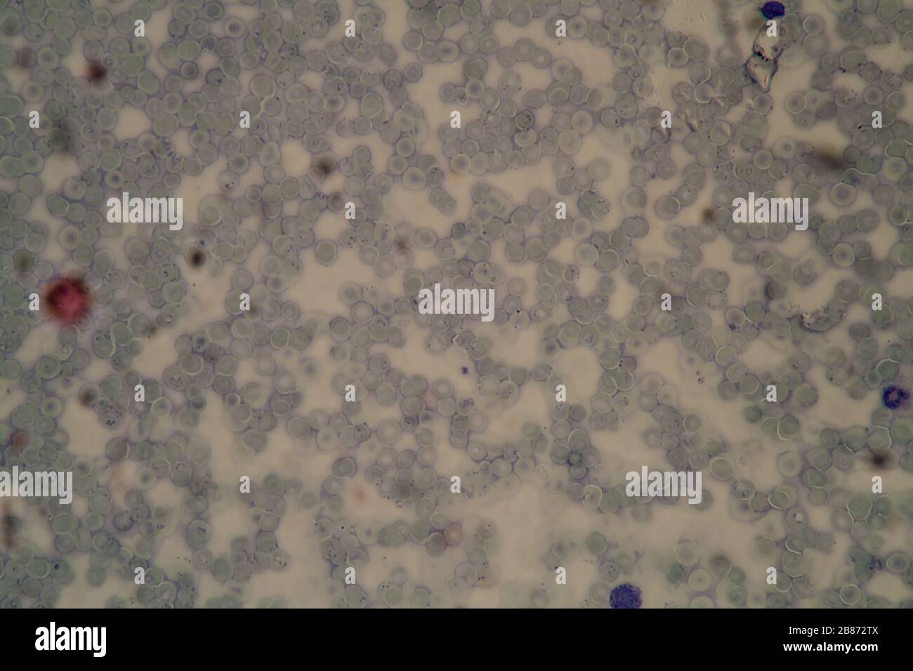
The malaria parasites in the ring trophozoites stage have size of about (1/5)th of the diameter of red blood cell.
Malaria parasite under the microscope view. Now, the same lensless microscope technique has been used to identify malaria parasites. Для просмотра онлайн кликните на видео ⤵. Malaria parasites of the genus plasmodium are diverse in mammal hosts, infecting five mammalian orders in the old world, but were long considered absent from the diverse deer family (cervidae) and from new world mammals. Hemozoin crystals are the byproduct of the malaria parasite, and they occur in the blood of an infected host. Detection of malaria parasites by light microscopy is still considered the primary method for malaria diagnosis in health clinics and hospitals throughout the the who practical microscopy guide for malaria provides detailed procedures for malaria diagnosis 6. The sporozoites which are slender. Utilizing machine learning algorithms, the microscope is so efficient that it can identify the amount of. Malaria is a serious disease caused by a blood parasite named plasmodium spp. Diagnosis depends on the quality of the stain and the expertise of the. Easyscan go, an ai powered microscope developed by chinese manufacturer motic, has the capability to automatically and accurately quantify malaria parasites in a blood sample.
Using a microscope, visual discovery. Can you find plasmodium parasites (malaria) in saliva under microscope from someone who's infected? Ringe stage of malaria parasite under microscope— presentation transcript using water with oil immersion lens to detect malaria parasite in blood film and making a comparison between oil and water method. The sporozoites which are slender. There was a description of a plasmodium parasite infecting a single.
Malaria parasite under the microscope view.
The malaria parasite is spread by female anopheles mosquitoes. Microscope shows malaria parasites in action. Malaria parasites present a tricky rare object problem for deep learning algorithms that typically require huge amounts of training data to accurately identify objects, says the tiny malaria parasites may only show up a handful of times within hundreds of microscope images of blood smears. Diagnosis of malaria involves identification of malaria parasite or its antigens/products in the blood of the patient. There was a description of a plasmodium parasite infecting a single. Malaria parasites of the genus plasmodium are diverse in mammal hosts, infecting five mammalian orders in the old world, but were long considered absent from the diverse deer family (cervidae) and from new world mammals. Malaria parasite under the microscope view. Automated method using microscope color image. Detection of malaria parasites by light microscopy is still considered the primary method for malaria diagnosis in health clinics and hospitals throughout the the who practical microscopy guide for malaria provides detailed procedures for malaria diagnosis 6. The malaria parasites in the ring trophozoites stage have size of about (1/5)th of the diameter of red blood cell.
The burst cells then infect the bloodstream via red blood cells, where the parasites reproduce further and eventually burst their host cell, leading to a vicious cycle. Diagnosis depends on the quality of the stain and the expertise of the. Malaria parasites take up giemsa stain in a special way in both thick and thin blood films. Malaria parasites invading human red blood cell подробнее. What we've achieved with mopid is the design of a polarized microscope platform using a cell phone, which can detect birefringence in histological specimens infected with the malaria. Easyscan go, an ai powered microscope developed by chinese manufacturer motic, has the capability to automatically and accurately quantify malaria parasites in a blood sample. Diagnosis plays a vital role in early detection compound light microscope, fluorescent microscopes, qbc centrifuge, refrigerator this prospective study was undertaken for detection of malaria parasite in 565 patients suffering. Place the glass slide on the microscope, with the label to the left. Utilizing machine learning algorithms, the microscope is so efficient that it can identify the amount of.

Malaria parasites take up giemsa stain in a special way in both thick and thin blood films.
The burst cells then infect the bloodstream via red blood cells, where the parasites reproduce further and eventually burst their host cell, leading to a vicious cycle. What we've achieved with mopid is the design of a polarized microscope platform using a cell phone, which can detect birefringence in histological specimens infected with the malaria. The malaria parasites in the ring trophozoites stage have size of about (1/5)th of the diameter of red blood cell. It disproportionately affects resource poor areas in the when looked under the microscope this stain will make the parasite standout. Utilizing machine learning algorithms, the microscope is so efficient that it can identify the amount of. Considering that malaria is a dreaded infection prevalent mostly in economically backward regions, an automated system for detection of malaria parasites in peripheral blood smear. Malaria parasite under the microscope view. Malaria parasites present a tricky rare object problem for deep learning algorithms that typically require huge amounts of training data to accurately identify objects, says the tiny malaria parasites may only show up a handful of times within hundreds of microscope images of blood smears. Diagnosis depends on the quality of the stain and the expertise of the. Ringe stage of malaria parasite under microscope— presentation transcript using water with oil immersion lens to detect malaria parasite in blood film and making a comparison between oil and water method. Для просмотра онлайн кликните на видео ⤵. David bell, director of global health technologies supporting global good. Using a microscope, visual discovery.
Malaria parasites take up giemsa stain in a special way in both thick and thin blood films malaria parasit. Conclusion the detection of malaria parasites is done by pathologists manually using microscopes.
 Source: onlinelibrary.wiley.com
 It disproportionately affects resource poor areas in the when looked under the microscope this stain will make the parasite standout.
 Source: www.researchgate.net
 Malaria parasites take up giemsa stain in a special way in both thick and thin blood films.
th of the diameter of red blood cell. Basic Malaria Microscopy Part I And Ii Learning Unit 8 Examining Blood Films For Malaria Parasites") Source: helid.digicollection.org
Source: helid.digicollection.org Malaria is one of the most important infectious diseases of humans and the most important parasitic disease.
 Source: c8.alamy.com
 The easyscan go uses custom image recognition software to identify and count malaria parasites in a blood smear in as little as 20 minutes.
 Source: journals.plos.org
 As the cause of both direct and indirect morbidity and mortality, it represents an enormous physical, psychic and also socioeconomic burden for the populations and states that it affects.
 Source: www.photonicsviews.com
 Diagnosis of malaria involves identification of malaria parasite or its antigens/products in the blood of the patient.
 Source: imgix.bustle.com
 Malaria parasite are transmitted by the bite of an infected female anopheles mosquito.
 Source: static.dw.com
 Malaria parasites of the genus plasmodium are diverse in mammal hosts, infecting five mammalian orders in the old world, but were long considered absent from the diverse deer family (cervidae) and from new world mammals.
 Source: www.researchgate.net
 The malaria parasite is spread by female anopheles mosquitoes.
 Source: helid.digicollection.org
 It disproportionately affects resource poor areas in the when looked under the microscope this stain will make the parasite standout.
 Source: c8.alamy.com
 The malaria parasites in the ring trophozoites stage have size of about (1/5)th of the diameter of red blood cell.
 Source: img.17qq.com
 Diagnosis depends on the quality of the stain and the expertise of the.
 Source: media.gettyimages.com
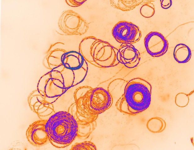
 Formally, the obtained spatial cellphone based microscope with a ball lens objective has been optimized for high resolution bright field imaging of malaria parasite in thin blood smears.
 Source: upload.wikimedia.org
 Place the glass slide on the microscope, with the label to the left.
 Source: d3i71xaburhd42.cloudfront.net
 The sporozoites which are slender.
 Source: images.freeimages.com
 Can you find plasmodium parasites (malaria) in saliva under microscope from someone who's infected?
 Source: blogs.biomedcentral.com
 Chandanmlt hello friends in this lecture explained about malaria life cycle/malaria parasite under microscope.
 Source: i2.wp.com
 The sporozoites which are slender.
 Source: www.isglobal.org
 Can you find plasmodium parasites (malaria) in saliva under microscope from someone who's infected?
 Source: i.pinimg.com
 Formally, the obtained spatial cellphone based microscope with a ball lens objective has been optimized for high resolution bright field imaging of malaria parasite in thin blood smears.
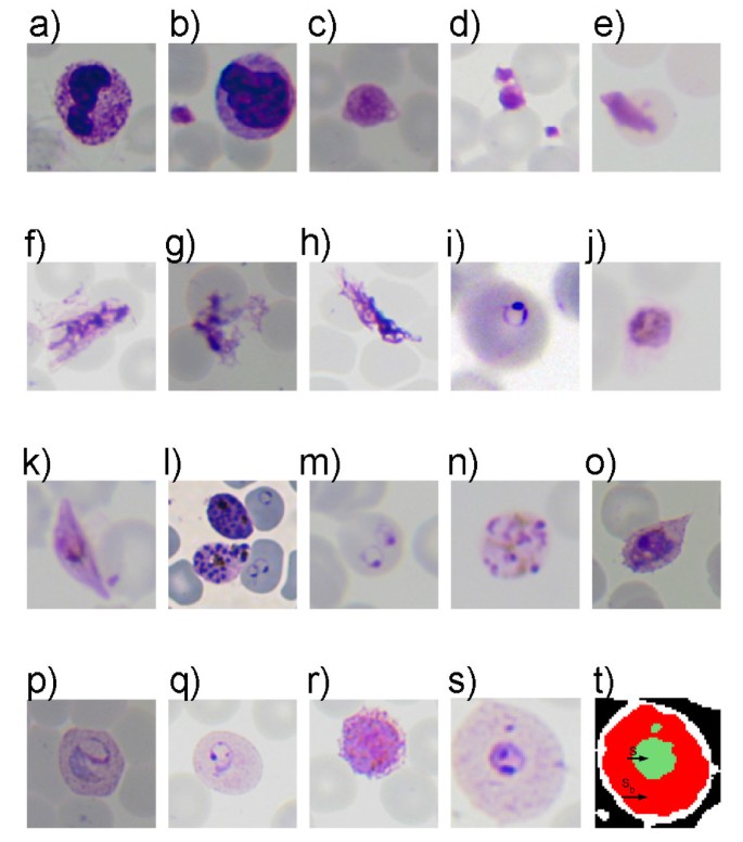
 and from new world mammals. Segmentation Of Erythrocytes Infected With Malaria Parasites For The Diagnosis Using Microscopy Imaging Sciencedirect") Source: ars.els-cdn.com
Source: ars.els-cdn.com The sporozoites which are slender.
Source: onlinelibrary.wiley.com Malaria is a serious disease caused by a blood parasite named plasmodium spp.
 Source: imaging.wehi.edu.au
 Malaria parasite are transmitted by the bite of an infected female anopheles mosquito.
 Source:
 The burst cells then infect the bloodstream via red blood cells, where the parasites reproduce further and eventually burst their host cell, leading to a vicious cycle.
 Source: image.shutterstock.com
 You should ask your tutor or facilitator to confirm the stage of parasite that you identify.
 Source: media.istockphoto.com
 Diagnosis of malaria involves identification of malaria parasite or its antigens/products in the blood of the patient.
 Source: images.freeimages.com
 Formally, the obtained spatial cellphone based microscope with a ball lens objective has been optimized for high resolution bright field imaging of malaria parasite in thin blood smears.
 Source: microscopy.anu.edu.au
 Performing a parasite count on a thick film and calculating parasite density 1.
 Source: www.mdpi.com
 Now, the same lensless microscope technique has been used to identify malaria parasites.
 Source: static3.bigstockphoto.com
 Formally, the obtained spatial cellphone based microscope with a ball lens objective has been optimized for high resolution bright field imaging of malaria parasite in thin blood smears.

Posting Komentar untuk "Malaria Parasite In Microscope"